
Artificial Intelligence-Integrated Radiographic Analysis for Developmental Dysplasia of the Hip Metrics in Infants and Children.
Abstract
Purpose:
Radiographic measures such as the acetabular index and IHDI classification are important metrics for diagnosing and monitoring developmental dysplasia of the hip (DDH). These measures are typically performed manually by radiologists and/or orthopedic surgeons, which can be time-consuming and introduce inter- and intra-rater variability. In this study, we introduce the preliminary development of an Artificial Intelligence (AI) system tailored for calculating DDH metrics to improve efficiency and reduce measurement variability.
Method:
We have utilized radiographs from a global prospective registry of infants and children diagnosed with DDH to develop an AI-integrated system. This proposed system combines two distinct components: an image segmentation model and a landmark detection model. Both of the image segmentation model and the landmark models are underpinned by the publicly available Segment Anything Model (SAM), notable for its efficiency with relatively small datasets.
Model Design: The design of the proposed model uses an assemble learning approach, as shown in Figure 1:

Figure 1: The Model Design
In the proposed model, both the segmentation model and landmark model employ pre-trained SAM models. By fine tuning the SAM model’s weights, the models are used for different tasks:
The segmentation model identifies different hip bone areas: the Ilium, capital femoral epiphysis and the rest of proximal femur bilaterally.
The landmark model is trained to learn the landmarks for DDH Metrics (triradiate cartilage and the superolateral edge of acetabulum).
We developed and evaluated the networks using AP pelvis radiographs obtained in 300 patients from a global prospective registry of patients with DDH with ethics board approval. In total 200 samples were used in the training process, 50 samples for testing and 50 samples for validation. Both of the pretrained SAM models finished the training in ~10 training epochs.
We compared accuracy of landmark localization vs. gold-standard human experts, the difference between the resulting AI-generated vs. expert-generated acetabular indices, and accuracy of IHDI classification (AI vs. expert via confusion matrix).
Results:
The preliminary result shows the proposed model converges quickly with relatively small samples to finish the training process. Figure 2a shows a predicted sample from the Segmentation Model and Figure 2b shows a predicted sample from the landmark detection model. These models will be used to automate measurement of the acetabular index and the IHDI grade.
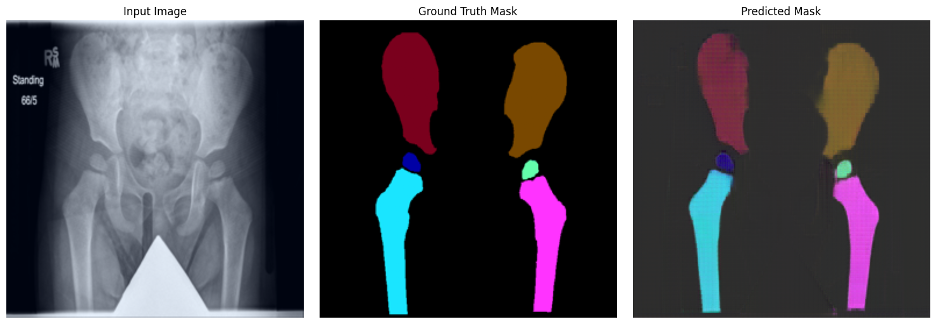
Figure 2a: Segmentation Prediction Sample

Figure 2b: Landmark Prediction Sample
Conclusion:
By integrating the outputs of these two models, we can utilize the identified landmarks for precise radiological measurements. Preliminary outcomes underscore the promising potential of our innovative approach. Effective AI-Integrated radiographic analysis will enable standardization of DDH metrics to improve research efficiency and comparability of results, while also holding potential to improve clinical efficiency and diagnostic accuracy, even in low-resource settings.
Disclosures:
Emily Schaeffer
Funded grants or clinical trials:
Orthopediatrics Pediatric Orthopaedic Society of North America Canadian Institutes of Health Research AO Foundation BC Children's Hospital Research Institute BC Children's Hospital Foundation